这个作者还真牛,各种照片很多。

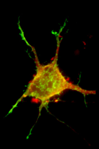
Here's a microscopy image of a fibroblast, stained with a few different antibodies. The green is microtubuli, the red is cell-contacts and the blue is DNA. It's just one of Jan Schmoranzer's amazing nano-art images.
Schmoranzer's microscopy images of "wounded monolayers," "starved fibroblasts" and a "nuclear face" come from the 2008-2009 NanoArt competition organized by NanoArt21.org.

Credit: DR JAN SCHMORANZER/SCIENCE PHOTO LIBRARY
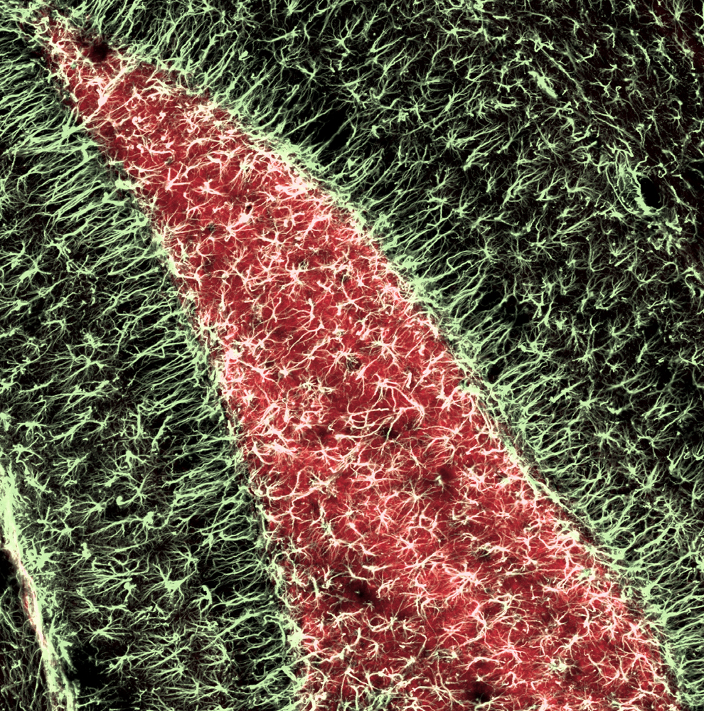
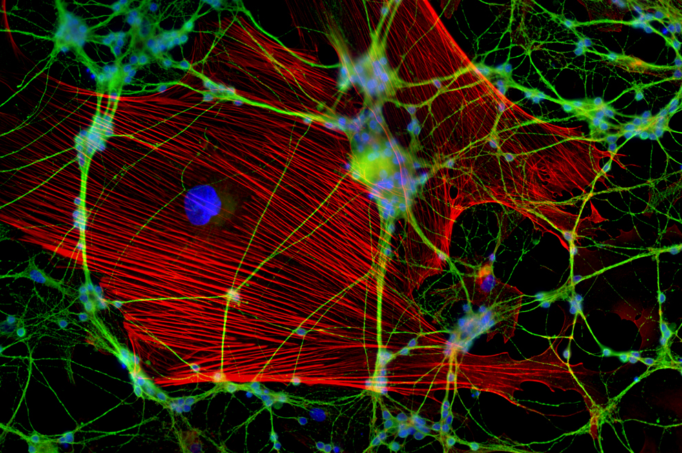
Caption: Glial cells. Immunofluorescence micrograph of glial cells. Glial cells are nervous system cells that provide structural support and protection for neurons (nerve cells). The cell nuclei, which contain the cells' genetic information, are blue. Microtubules are green and actin filaments are red. These protein filaments make up part of the cytoskeleton, which maintains the cells' shape, allows some cellular mobility and is involved in intracellular transport. Magnification: x3200 when printed at 10 centimetres wide.